Current Research Projects
Snake Muscle Evolution
Snakes rely on extensive cranial kinesis to swallow large prey without the aid of limbs. This has reached its apex in the "pterygoid walk", where snakes "walk" their heads over their prey by unilaterally advancing their palate and mandibles. For this project I am investigating the musculature that powers cranial kinesis in snakes using 3D DiceCT data. With this data, I aim to understand how muscular morphology relates snake feeding behavior and evolution.

Fascicle models of the palatal muscles in a rough scaled death adder, Acanthophis rugosus
Fiber Tracking Methods
New methods for fiber tracking have been applied to measure fibrous biological tissues such as cartilage and muscle. I have am currently building on these methodologies for usage on DiceCT and other 3D biological data. This includes developing methods and protocols for measuring muscle architecture with fiber tracking software and comparing the results of existing tools. The muscle architectural measurements derived from these tools can be applied to studies of anatomy, evolution, and biomechanics of diverse musculoskeletal systems.

Avizo Xfiber and Goodfibes models of the temporalis of a Virginia opossum
Biomechanics and Anatomy of Macrostomy
Macrostomy, or enlarged gape, is a key innovation of snakes that allows for specialization on bulky prey items. While we have a good understanding of the bony structure that allow for expanded gape, data on how soft tissue structure survive these stresses is much more limited. For the second chapter of my dissertation, I am using DiceCT to investigate the effects of gape on the biomechanics and architecture of snake jaw muscles.

DiceCT data of a Florida Watersnake (Nerodia fasciata pictiventris) at maximum gape
Pangolin Tongues as Muscular Hydrostats
Pangolins tongues are highly derived structures that extend nearly a body length to catch food, and yet are fully packed into the thoracoabdominal cavity at rest. Using DiceCT, I am describing the anatomy of the tongue and hyolingual apparatus of the white-bellied pangolin to understand the mechanics of this complex feeding structure. Descriptions of the 3D fascicle architecture of a portion of the pangolin tongue support its function as a muscular hydrosat, recently published in Integrative & Comparative Biology.
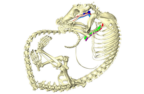
CT data of a white-bellied pangolin showing the skeleton and select portions of the tongue and oral cavity
Fanged Frog Morphology
The frogs of the genus Limnonectes are known as the "fanged frogs" for the large odontoid "fangs" on their mandibles. In collaboration with Dr. Johana Goyes Vallejos, we are looking into the mandible morphology of members of the genus using micro-computed tomography to study the shape and allometry of these fangs in relation to ecology and behavior.
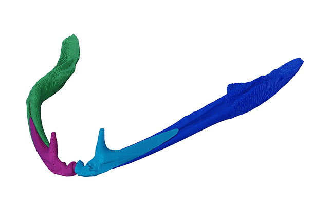
Segmented mandible of Limnonectes magnus